The Radioactive Dinner that Started Nuclear Medicine
When people hear "nuclear medicine" they tend to think of something that's straight out of a Cold War movie, but it's actually one of the quietest, most useful corners of modern medicine. Every year, millions of people get a scan or a treatment that relies on a tiny amount of radioactive material, and most of them never think twice about it.
To see how we got here, it helps to start with a suspicious landlady, a question about iodine, and a patient named Elizabeth.
Step 1: Radioactivity becomes a tracer (1890s to 1930s)
Radioactivity was discovered in the 1890s and Marie and Pierre Curie's work on radium made it famous, with the early medical idea being simple enough: radiation kills cells, so point it at tumours.
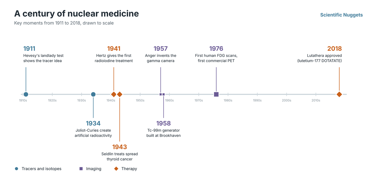
The bigger idea came from a young Hungarian chemist named George de Hevesy, and the story goes that around 1911, while he was working in Ernest Rutherford's lab, Hevesy suspected the landlady of his Manchester boarding house of recycling the week's leftover meat into later meals. She denied it, so Hevesy secretly sprinkled a tiny amount of radioactive material onto the leftovers on his plate and a few days later brought an electroscope, an early radiation detector, to dinner and showed that the "fresh" meal was radioactive. Hevesy never published the episode himself and the details vary between retellings, but it appears in the standard histories of nuclear medicine (see sources at the end).
The lesson behind the prank is real, because a trace amount of a radioactive atom behaves chemically like its stable twin and a detector can follow it wherever it goes. It's a bit like sewing a tiny GPS beacon into a molecule. Hevesy went on to use radioactive lead to track how plants move lead through their tissue in 1923 and won the 1943 Nobel Prize in Chemistry for the tracer principle. He is often called the father of nuclear medicine, and that same idea is behind every PET tracer today, including the PSMA scan in my last post.
The catch was that back in the day, the only radioactive materials available were rare natural ones, but that changed in 1934 when Irène and Frédéric Joliot-Curie (the daughter and son-in-law of Marie and Pierre) showed that radioactive versions of ordinary elements could be made in the lab. The right machine already existed to do just that as Ernest Lawrence, an American physicist, had a cyclotron at Berkeley that could produce radioactive isotopes on demand, meaning scientists suddenly had a whole catalogue of beacons to choose from.
Step 2: The iodine question (1936 to 1946)
The thyroid made some of the first radioactive drugs possible, because it pulls iodine out of the blood to make its hormones, so radioactive iodine can go straight to the gland without any chemistry to steer it there. Nothing else in the body offers such a convenient built-in target.
In November 1936, a Boston physician named Saul Hertz attended a Harvard Medical School faculty luncheon where MIT president Karl Compton was speaking about "What Physics Can Do for Biology and Medicine." After hearing this, Hertz asked Compton whether iodine could be made radioactive, and a month later MIT's president wrote back that it could. Hertz then teamed up with physicist Arthur Roberts to find out if the thyroid would take up radioiodine just as it takes up ordinary iodine, and they started with rabbits. The iodine they could make at MIT lost half its radioactivity in only 25 minutes, which was too short-lived to treat anyone, so the move to people had to wait until MIT's new cyclotron began running in late 1940 and produced a longer-lasting form.
On March 31, 1941, Hertz gave radioactive iodine to his first human patient named Elizabeth who had an overactive thyroid, and over the next two years he treated 29 patients, of whom 20 were considered cured. This is considered one of the first examples of targeted therapies: a drug that finds its own way to the diseased tissue and treats it from the inside.
In 1943, Samuel Seidlin, an endocrinologist at Montefiore Hospital in New York, gave radioactive iodine to a patient whose thyroid cancer had spread, and his 1946 report on that patient's response is widely considered the paper that put nuclear medicine on the map, while Hertz, who died young in 1950, got far less credit than he deserved for decades.
The isotope behind all of this, iodine-131, had been created at Berkeley in 1938 by Glenn Seaborg and John Livingood, and the idea at its core turned out to be bigger than the isotope itself, because the same element was being used both to see a disease and to treat it.
Step 3: The workhorse arrives (1950s to 1960s)
Iodine-131 worked well for treatment but was not ideal for imaging, since it lingers for about eight days and gives patients a larger radiation dose than you would want from a routine scan.
The fix was technetium-99m, a form of an element that has no stable isotopes and is essentially absent from nature. It was the first element to be made artificially and it was named after the Greek word for artificial. Emilio Segrè and Carlo Perrier first isolated it in the late 1930s from a piece of molybdenum that had been bombarded in Lawrence's cyclotron, while they were hunting for the missing element 43 on the periodic table, and its properties turned out to be almost perfect for imaging. It had a half-life of about six hours and a clean gamma ray that a camera can see easily. A generator developed at Brookhaven in 1958 made it practical to produce at hospitals, and Powell Richards, who ran isotope production there, began promoting it as a medical tracer in 1960, after which it became the most used isotope in nuclear medicine, and it is still involved in a large share of nuclear imaging scans today.
The other piece was the camera, because in 1957 Hal Anger developed the gamma camera that turned scattered radiation signals into pictures of organs at work, and by the early 1970s nuclear medicine was officially recognized as its own medical specialty.
Step 4: PET and the sugar trick (1970s)
If Tc-99m shows you where something is, PET shows you what it’s doing.
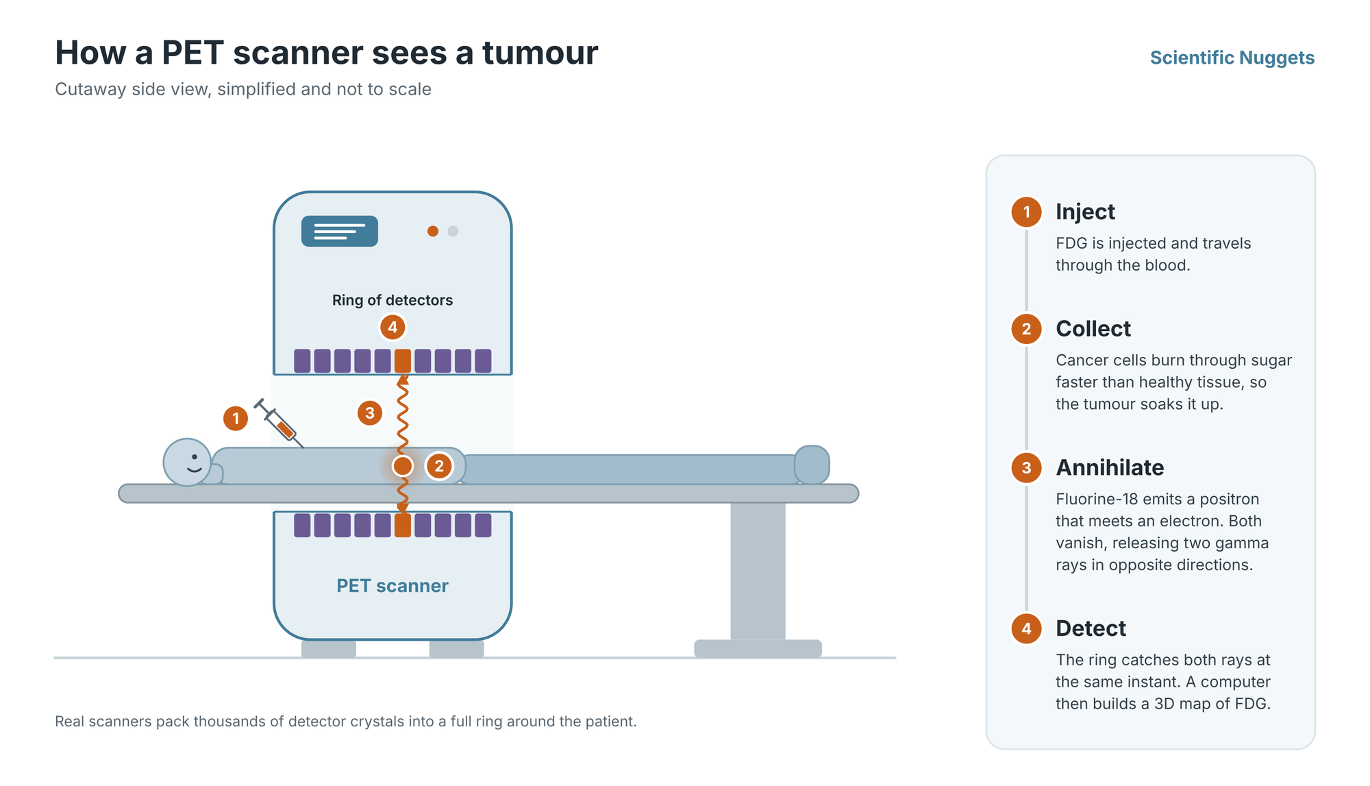
Positron emission tomography (PET) was built in the 1970s by Michel Ter-Pogossian, Michael Phelps, and colleagues at Washington University in St. Louis. Their scanner detected the pairs of gamma rays released when a positron meets an electron, and a computer reconstructed a 3D map from them. The first commercial PET scanner was delivered to UCLA in 1976.
The companion breakthrough was a radioactive sugar called FDG, short for fluorodeoxyglucose, made by tagging a glucose analogue with radioactive fluorine-18. Cancer cells burn through sugar faster than most healthy tissue, so a tumour lights up like a flare. The first human FDG images were taken in 1976 at the University of Pennsylvania, using material made at Brookhaven National Laboratory. Half a century later, FDG is still the most widely used PET tracer in the world.
It took roughly two decades for PET to move from a clever prototype to a routine clinical tool, with funding and technology being the bottleneck rather than the idea.
Step 5: Back to iodine's big idea (2000s to now)
For most of the 20th century, nuclear medicine ran on two tracks. Diagnosis used one set of isotopes, and therapy used another. Over the last 25 years, those tracks have merged into what the field calls theranostics: a pair of radiopharmaceuticals that share the same targeting molecule. One carries an imaging isotope to show whether the target is there. The other carries a therapeutic isotope to destroy it.
This is Hertz's iodine story scaled up with modern chemistry. Gallium-68 DOTATATE (approved in the US in 2016) lets doctors see neuroendocrine tumours on a PET scan, and its therapeutic partner, lutetium-177 DOTATATE (approved in 2018), treats them. In prostate cancer, the same logic now underpins Pluvicto, which was the subject of my last post.
What the history tells us
Looking back, the breakthroughs came from chemists, physicists, and physicians asking each other naive questions, and every leap forward from iodine to technetium to fluorine-18 to lutetium happened because someone found a new isotope or a new way to attach one to a molecule that goes where you want it to. This is why the next chapter is being written right now in labs by people like me who are designing new tracers from scratch.
Sources
- Hertz and the first radioiodine treatment: Hertz S, EJNMMI Physics review
- Overview of Hertz's work: Hertz, Intechopen chapter
- Technetium-99m and I-131 history: PMC3339145
- Hevesy and the tracer principle: J Nucl Med Technol, Myers
- Berkeley Lab isotope discoveries: Berkeley Lab
- First human FDG imaging and approvals: SNMMI, J Nucl Med 58(1)
- PET history: Phelps, DOE Fermi Award
- Hevesy and the landlady story (retold from Myers 1979 and Brecher and Brecher 1969): Four Tales of George de Hevesy, ORAU Health Physics Museum
- Published account of the anecdote (secondhand, since Hevesy never wrote it up): Myers WG, J Nucl Med 1979;20:590-594